Wednesday, August 27, 2014

Giant Deep Sea Amoebas! Meet the Xenophyophores!!

So, following up with all of the NOAA deep-sea Okeanos Explorer stuff, I've found that I am just FASCINATED by these things called Xenophyophores!! The name means "Bearer of foreign bodies".

But what are they? To put it they simply, they are giant, deep-sea amoebas that live in large, sediment "houses" called "tests" (similar to the way that echinoderm skeletons are also known as tests).

Footnote on the classificaiton: a quick survey of the various Protist classifications tells me that even calling these organisms "amoebas" is probably incorrect. But the nuances of this dynamic are for another day..

This one group, the Xenophyophorea live in the deep-sea.. DEEP in the deep-sea! Xenophyophores were observed as deep as10 KM (over 6 miles!) in the deepest of marine trenches (the Mariana) and occur in almost ALL of the world's oceans (except the Arctic).

They are considered among the world's largest living SINGLE CELLED organisms.

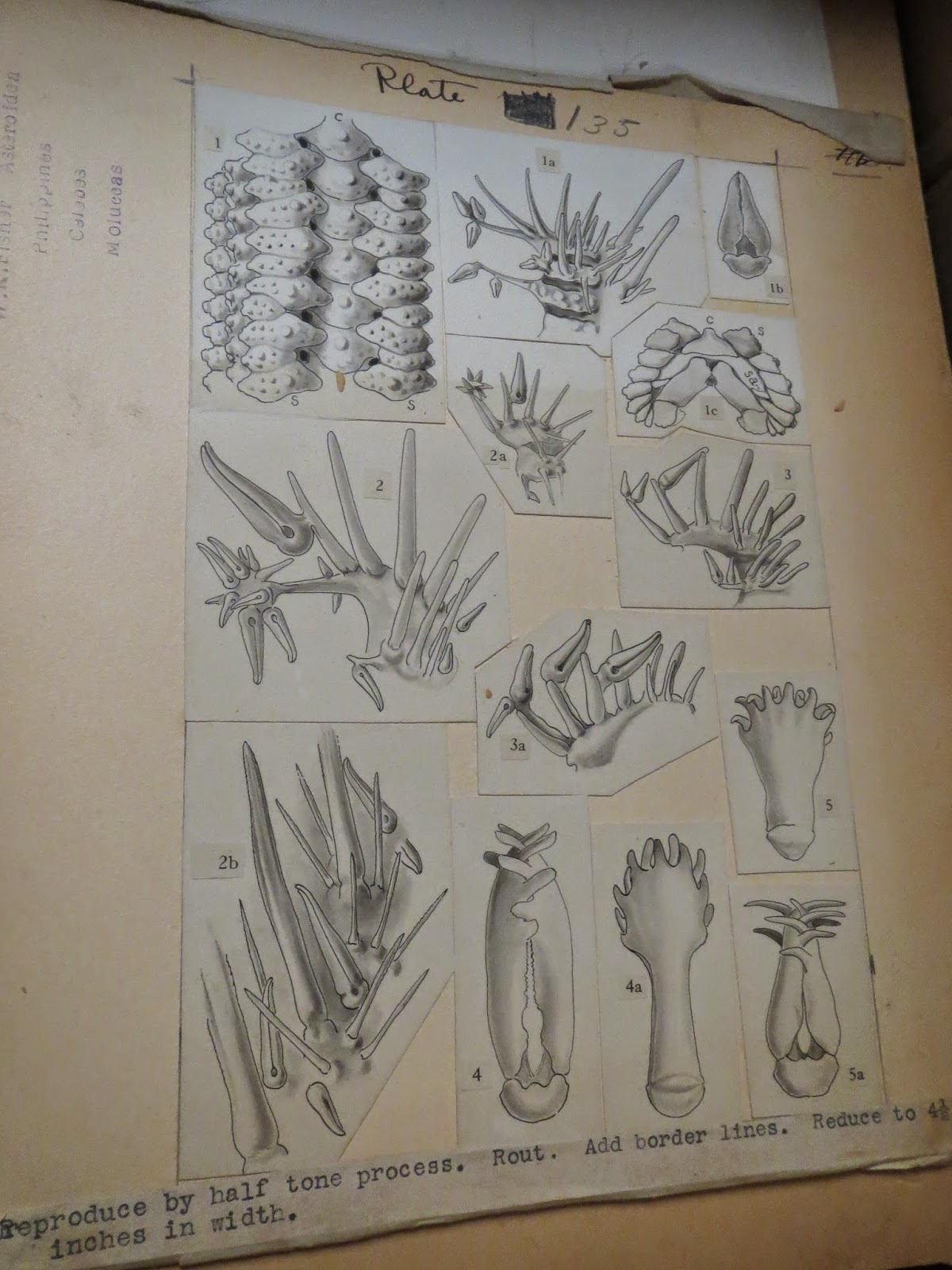
Xenophyophores create these large tests which they inhabit. The tests come in a variety of forms. There are 42 known species in 13 genera. As I understand it, xenophyophores are considered as a subgroup within the Foraminifera (these are amoeba-like unicellular organisms with tests).

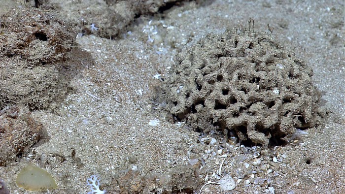
Here's a variety of xenophyophore tests spied by the Okeanos Explorer below..but other patterns include big leafy structures and more network-like arrays of tubes. They vary quite a bit...
                              From the NOAA Photo Library here.                            
From the NOAA Photo Library here
From the Gulf of Mexico, NOAA photo library
Here's some fun facts!  Several different sources including the "Paleoecology and ecology of Xenophyophores" by Lisa Levin and others cited below...

1. How Big Are They?? So, here's the thing. I've read plenty of accounts that jump to the conclusion "These are the MONSTER AMOEBAS!!!" But what a lot of these accounts seem to forget is that the large sizes considered by a lot of popular accounts are the TESTS (i.e., the skeletons).

To be sure, these structures can be pretty big (for something made by amoebas!) The example below is apparently about 25 cm (almost a foot!) across!

Most of the accounts I've read sort of assume (and I suppose this is reasonable) that the animal inhabits ALL of the test all the time, or perhaps with pseudopods or tentacles extended throughout? Frankly, none of the accounts I read could clarify how much of the test, the actual organism inhabits.

However, One estimate (here) indicates that the test volume might be as little as < 1% "protoplasm" (which if I understand the terminology includes cytoplasm, etc.).. so, the actual organisms are probably not as monstrous as some folks would think. I would imagine its quite difficult to measure an amoeba for something like this.


2. What Do They Eat?
  So, when you think of big deep-sea amoebas, perhaps automatically we think "oh WOW! Wouldn't it be NEAT if they actually could eat ANIMALS?" Just like in the movies??  And in truth, there ARE marine amoebas which probably devour animals ... but to date, very little evidence is available on the full range of what xenophyophores actually eat. and the truth is sadly not likely to be as romantic as some would think...
One paper by Laureillard, Mejanelle and Sibuet from 2004 studied the xenophyophore Syringammina corbicula and utilized a study of various lipids and amino acids to look at their nutrition. Their study showed that bacteria were present in great abundance!

Xenophyophores have strings of mucus which are deployed along the test which build up feces and sediment called stercomes. It was suggested that a flora of these bacteria were present in abundance on these mucous threads. Perhaps being farmed and being utilized as a source of food.

Their mucous threads also are constantly pulling and trapping particles from the surrounding area, presumably in part to provide further nutrition.

Here is an Scanning Electron Microscope Image of a stercome showing up close details of what's on them...

So, there seems to be a heavy dependence on poop and other "marine snow" that falls down to the bottom.. as well as bacterial/microbial growth. But very little is known about feeding in xenophyophores, so who knows what else they do??

3. What are those structures (tests) made of? 
Tests on xenophyophores are made up of a patchwork of different bits. Sediment, but also the shells of other marine organisms such as radiolarians, other foraminiferans, and so on...

The test is the outer "crunchy" later... Within the test are a series of tubes called granellare, through which the animal's cytoplasm and etc. flow through...

These tests are actually an important part of xenophyophores ecological role, as they provide habitats and such.....

4. Ecology!
Probably the most interesting thing that I've picked up about Xenophyophores?? Is how potentially important they are to deep-sea ecosystems. Xenos are VERY abundant in the deep-sea, sometimes reaching up to 2000 per 100 square meters!

Lisa Levin published this neat paper in 1991 about their roles in deep-sea communities.  Basically it turns out that where xenos are found, there are "hotspots" of animal diversity!

The pic below shows two big xenophyophore tests with brittle stars on them...
Why might a cluster of xenophyophore tests represent a "hotspot" of animal diversity?? They apparently present both food AND habitats for a plethora of deep-sea animals!

Examples of how animals use these tests?
  • Habitats for worms, copepods, crustaceans, ophiuroids, and even snail embryos!
  • Peanut worms that live IN "dead" tests
  • Eggs from various animals (worms or snails) on xenophyophore tests.
  • Suspension feeding colonial animals called bryozoans sometimes are found "intergrown" on tests
  • Some xenophyophores found ON sea urchins.
  • Some amphipod crustaceans are thought to prey on the xenophyophores
  • Plus other MORE! Possibly/probably associated with the bacteria growing on the surface?? 
5. Mystery fossils: Xenophyophores??
So, one of the aforementioned articles by Levin on Xenophyophore paleoecology makes the case that tests and other modern examples of sediment structures by these organisms can explain various trace fossils, and other mysterious fossil structures.

Perhaps one of the best known is that of the fossil ichnogenus (like an organism name but for a trace fossil) Paleodictyon!

Paleodictyon was famously studied by oceanographer Peter Rona who made it an obsession of his career. It has covered by many other natural history blogs such as this and this. 

Basically.. a trace fossil which looks like this.
This is seen throughout the deep-sea in many places. The above image from the Gulf of Mexico but its been seen throughout the deep-sea, sometimes as deep as 3700 meters!!

Its also been seen in the fossil record.. dating back to the Paleozoic...  The parallel and similar appearance has led to much debate over whether the same type of organism has created these patterns in the sediment.. namely, did a xenophyophore or something similar create them?? 
And anyway, there's a LOT more on them in this regard and there seem to be more questions than answers.... A neat group of critters! (can I use that for amoebas?) 

Wednesday, August 20, 2014

Golden Tickets in the NOAA Photo Library! Rarely seen Pelagic Sea Cucumber!

From NOAA Photo Library here. 
HOLY CARP!! So, as you know, I've been going through and finding some GREAT stuff in the NOAA Photo Library (see this post from a few days ago) but every once in awhile you find one that is especially... striking!

This is the "golden ticket" so to speak.. from Charlie & the Chocolate Factory! that moment when Charlie finds that rare golden ticket from millions of chocolate bars! That special ticket to the rare tour of the chocolate factory! From MANY, MANY hours of going through these pictures I've found several GREAT items.
To be sure, there were quite a few "golden tickets" to be found among the thousands of pictures in the NOAA Photo Library (and for professional reasons I haven't shared all of them), but this one made me especially excited!!!   Why??

Because this is probably one of the FIRST public images of the "proper" Swimming sea cucumber Pelagothuria, possibly Pelagothuria natatrix!!  See my post here.  Dr. Dave Pawson at the NMNH confirms its identity. The image was taken from the Galapagos Rift Expedition in 2011. So its been sitting around for several years! 

Translation: A TRUE SWIMMING SEA CUCUMBER and probably the ONLY swimming (i.e. pelagic) echinoderm known!!! 
From NOAA Photo Library here. 
What you're seeing above is the swimming "umbrella" around the mouth, which is facing upwards! The body is the cone -shaped bit below it. 

This species occurs between 570 and 6000 meters in the Atlantic, Pacific and Indian Oceans but is poorly known. 

Previously.. this animal was known only from pictures....
Or from these grainy videos....(from Miller & Pawson)
 I have no doubt that there's scientists (my colleagues) out there who have seen this before and probably have video of this species..but this is the first that is available to share with ANYONE!!

So that's why you guys are getting TWO posts this week!!

Tuesday, August 19, 2014

Because Brisingid Starfish are Fantastic! Stunning Images of Brisingids in the Deep!

This image from NOAA Photo Library. Here. 
Brisingid starfishes were my first professional "love".. I wrote my Masters thesis on them and it was almost 2 years after studying specimens like this...
Before I saw one that was alive!  Now, THAT is dedication for you! 

What are brisingid starfish?? Long story short... Strange deep-sea starfish. They are proper, albeit highly modified sea stars that use their spines, covered with tiny claws which act as velcro to capture tiny prey as food.
This is Novodinia. Photo form NOAA Okeanos Explorer

I was just noticing that there's been a great critical mass of REALLY wonderful pictures of these animals.

For example, here's an amazing close up shot of Novodinia, possibly N. americana from the R/V Okeanos Explorer cruise to the North Atlantic canyons in 2013.  What you're seeing is the disk at the center, surrounded by the many spines covering each of its arms...
This image originally from NOAA Photo Library here. 
shots like these are increasingly common..but trust me when I say that scientists from the mid late 20th Century would have KILLED to have nice high definition picture like this!
Another Pic of Novodinia americana? from 2013. NOAA Photo Library. 
And here was one AMAZING bit of anecdotal observation/biology from the 2013 North Atlantic Okeanos Explorer cruise, this brisingid, unclear which genus, based purely on the pic not only caught a fish but HELD onto it using ONLY its pedicellariae.
Owly Images
Pedicellariae are tiny claw-shaped structures that cover each of those spines..sort of like staples embedded in a sock. These capture various food and prey items.. but mostly it was thought they captured crustaceans. Capturing fish is a bit unusual....

Food caught by the spines and on the surface are then moved down to the underside to the mouth...

Colleague Jackson Chu, provides us with a GREAT pic of the UNDERSIDE of a brisingid, showing the mouth, tube foot grooves and etc.. just what you would expect from any proper sea star...
                    

Here are some stunning panoramic shots of brisingids. Presumably, these occur on places where water currents are favorable for them to capture food...  Both of these are from the North Atlantic via Okeanos Explorer..

These animals feed by holding their arms up into the water and capturing food/prey as it is carried by on the water currents...

This shot was from a spur projecting from the canyon wall in the North Atlantic (Block Canyon) in 2062-2131 m. 

This pic from 2013 Atlantic Canyons Expedition
Here are some great shots from Neptune Canada via Flickr...  A large individual, maybe Brisinga? on a sunken barrel..
                    
some very "at attention" individuals...
                          
Not all species occur on hard bottoms.. Some live on mud and sediment...
This one from the NE Canyons expedition in 2013
And just for a little diversity, From Japan, here is the underside of what I think is Brisingaster or Novodinia... These likely represent an unpublished record of this species in Japan..

Here is what the top side looks like...

Tuesday, August 12, 2014

5 Cool, Weird Brittle Star Behavior Captured on video/pictures!

Ophiuroids (aka serpent stars and brittle stars) do some pretty remarkable things, BUT They are often tiny and cryptic so we don't get to see the cool array of things they do.

Flickr and YouTube have provided us with a HUGE quantity of imagery of interesting aspects of ophiuroid biology. Here is a sampling of the ones I found worth sharing!

1. Swollen Disk! This is the deep-sea serpent star Asteronyx! which live with arms wrapped around deep-sea cnidarians, such as sea pens. But this one has a fully inflated disk! When we study them in the lab, these are deflated and flattened. What do they do with them when inflated?  Why? 

This one is probably a different species, but it gives you an idea of how these live...with their arms wrapped around a cnidarian called a sea pen..


2. Brittle Star Burrowing!?  See those two brittle star arms emerging from the burrow?? That means the disk is buried within the sediment. But WOW! Look at the sediment being dumped out of that burrow in a continuous string! Maybe something else is in there putting that out? or is the brittle star doing that??


One of the rays of an amphiurid. Note the tube feet/spines fully occupied by sediment as it digs its way into the bottom.


That arm is probably part of this critter.. an amphiurid brittle star


3. Brittle Stars Feeding: Tube feet in ACTION!! How often do you get to see a brittle star in full feeding action??


and even MORE ophiuroid feeding action!!!


4. Brooding Brittle Star CT SCAN! Here is an internal CT scan of a BROODING ophiuroid! The ones inside the disk are juveniles!


5. Brittle Star "SWIMS"! This looks like Ophiocnemis, the brittle star which seems to find itself hitching a ride in jellyfishes! I've written about these here! 

Tuesday, August 5, 2014

Before Computers! Deep-Sea Starfish Plates from circa 1919!


Something a little different today! There have been some plates from old 20th Century starfish & other echinoderm monographs in storage in and around our lab space and they are just gorgeous!  I've shared some of these before (several years ago here) But there are MANY of them....


Image from this MBL page
Fisher wasn't perfect, but he is considered one of the giants of starfish taxonomy and described about 312 species of starfishes that continue to remain in use. He also described sea cucumbers, peanut worms, stylasterine corals and probably more.

Much of his work was done as huge monographic tomes which included hundreds of pages of scientific descriptions including some of the finest photographic figures available. 
And he did this way before computers. No word processing. No internet. 
Before we had photoshop and imaging software, publishing was a very physical process. Photographs were mounted to cardboard as seen below. So, the "plates" were quite literally so. These got quite big and cumbersome.
No photoshop, so images were pasted together by hand as so..
Here is a plate from an ophiuroid or brittle star plate from a monograph by French researcher Rene Koehler. Also assembled from a photograph.
                                 
If you wanted to modify or target images for plates, there was no image modification software to do it for you, you literally had to take an exacto knife or razor blade to photographs, cut them out and affix them to the hardboard plates...
                  
But the images were done in amazingly high quality and remain attractive to this day.. Here are scans of the ORIGINAL plates from Fisher's Philippine Starfish monograph. No Instagram. No other photo modification.  These are directly off the original plates..


some from before...